 Your new post is loading...
Can knowing treatment options for a dislocated hip help individuals expedite rehabilitation and recovery? Dislocated Hip A dislocated hip is an uncommon injury but can happen due to trauma or following hip replacement surgery. It usually occurs after severe trauma, including motor vehicle collisions, falls, and sometimes sports injuries. (Caylyne Arnold et al., 2017) A dislocated hip can also occur after hip replacement surgery. Other injuries like ligament tears, cartilage damage, and bone fractures can occur alongside the dislocation. Most hip dislocations are treated with a joint reduction procedure that resets the ball into the socket. It is usually done with sedation or general anesthesia. Rehabilitation takes time and could be a few months before full recovery. Physical therapy can help restore motion and strength in the hip. What Is It? If the hip is only partially dislocated, it's called a hip subluxation. When this happens, the hip joint head only partially emerges from the socket. A dislocated hip is when the head or ball of the joint shifts or pops out of the socket. Because an artificial hip differs from a normal hip joint, the risk of dislocation increases after joint replacement. A study found that around 2% of individuals who undergo total hip replacement will experience hip dislocation within a year, with the cumulative risk increasing by approximately 1% over five years. (Jens Dargel et al., 2014) However, new technological prosthetics and surgical techniques are making this less common. Hip Anatomy - The hip ball-and-socket joint is called the femoroacetabular joint.
- The socket is called the acetabulum.
- The ball is called the femoral head.
The bony anatomy and strong ligaments, muscles, and tendons help to create a stable joint. Significant force must be applied to the joint for a hip dislocation to occur. Some individuals report feeling a snapping sensation of the hip. This usually is not a hip dislocation but indicates a different disorder known as snapping hip syndrome. (Paul Walker et al., 2021) Posterior Hip Dislocation - Around 90% of hip dislocations are posterior.
- In this type, the ball is pushed backward from the socket.
- Posterior dislocations can result in injuries or irritation to the sciatic nerve. (R Cornwall, T E Radomisli 2000)
Anterior Hip Dislocation - Anterior dislocations are less common.
- In this type of injury, the ball is pushed out of the socket.
Hip Subluxation - A hip subluxation occurs when the hip joint ball starts to come out of the socket partially.
- Also known as a partial dislocation, it can turn into a fully dislocated hip joint if not allowed to heal properly.
Symptoms Symptoms can include: - The leg is in an abnormal position.
- Difficulty moving.
- Severe hip pain.
- Inability to bear weight.
- Mechanical lower back pain can create confusion when making a proper diagnosis.
- With a posterior dislocation, the knee and foot will be rotated towards the body's midline.
- An anterior dislocation will rotate the knee and foot away from the midline. (American Academy of Orthopaedic Surgeons. 2021)
Causes A dislocation can cause damage to the structures that hold the ball in the socket and can include: - Cartilage damage to the joint -
- Tears in the labrum and ligaments.
- Fractures of the bone at the joint.
- Injury to the vessels that supply blood can later lead to avascular necrosis or osteonecrosis of the hip. (Patrick Kellam, Robert F. Ostrum 2016)
- A hip dislocation increases the risk of developing joint arthritis following the injury and can raise the risk of needing a hip replacement later in life. (Hsuan-Hsiao Ma et al., 2020)
Developmental Dislocation of the Hip - Some children are born with developmental dislocation of the hip or DDH.
- Children with DDH have hip joints that did not form correctly during development.
- This causes a loose fit in the socket.
- In some cases, the hip joint is completely dislocated.
- In others, it's prone to becoming dislocated.
- In milder cases, the joint is loose but not prone to becoming dislocated. (American Academy of Orthopaedic Surgeons. 2022)
Treatment Joint reduction is the most common way to treat a dislocated hip. The procedure repositions the ball back into the socket and is usually done with sedation or under general anesthesia. Repositioning a hip requires significant force. A hip dislocation is considered an emergency, and reduction should be performed immediately after the dislocation to prevent permanent complications and invasive treatment. (Caylyne Arnold et al., 2017) - Once the ball is back in the socket, the healthcare provider will look for bone, cartilage, and ligament injuries.
- Depending on what the healthcare provider finds, further treatment may be necessary.
- Fractured or broken bones may need to be repaired to keep the ball within the socket.
- Damaged cartilage may have to be removed.
Surgery Surgery could be necessary to return the joint to its normal position. Hip arthroscopy can minimize the invasiveness of certain procedures. A surgeon inserts a microscopic camera into the hip joint to help the surgeon repair the injury using instruments inserted through other small incisions. Hip replacement surgery replaces the ball and socket, a common and successful orthopedic surgical procedure. This surgery may be performed for various reasons, including trauma or arthritis, as it is common to develop early arthritis of the hip after this type of trauma. This is why many who have a dislocation ultimately need hip replacement surgery. As a major surgical procedure, it is not without risks. Possible complications include: - Infection
- Aseptic loosening (the loosening of the joint without infection)
- Hip dislocation
Recovery Recovering from a hip dislocation is a long process. Individuals will need to walk with crutches or other devices early in recovery. Physical therapy will improve the range of motion and strengthen the muscles around the hip. Recovery time will depend on whether other injuries, such as fractures or tears, are present. If the hip joint was reduced and there were no other injuries, it may take six to ten weeks to recover to the point where weight can be placed on the leg. It could be between two and three months for a full recovery. Keeping weight off the leg is important until the surgeon or physical therapist gives the all-clear. Injury Medical Chiropractic and Functional Medicine Clinic will work with an individual's primary healthcare provider and other surgeons or specialists to develop an optimal personalized treatment plan. General Disclaimer * The information herein is not intended to replace a one-on-one relationship with a qualified healthcare professional or licensed physician and is not medical advice. We encourage you to make healthcare decisions based on your research and partnership with a qualified healthcare professional. Our information scope is limited to chiropractic, musculoskeletal, physical medicines, wellness, sensitive health issues, functional medicine articles, topics, and discussions. We provide and present clinical collaboration with specialists from various disciplines. Each specialist is governed by their professional scope of practice and their jurisdiction of licensure. We use functional health & wellness protocols to treat and support care for the injuries or disorders of the musculoskeletal system. Our videos, posts, topics, subjects, and insights cover clinical matters, issues, and topics that relate to and directly or indirectly support our clinical scope of practice.* Our office has reasonably attempted to provide supportive citations and identified the relevant research studies or studies supporting our posts. We provide copies of supporting research studies available to regulatory boards and the public upon request. We understand that we cover matters that require an additional explanation of how it may assist in a particular care plan or treatment protocol; therefore, to further discuss the subject matter above, don't hesitate to contact Dr. Alex Jimenez or contact us at 915-850-0900. Dr. Alex Jimenez DC, MSACP, CCST, IFMCP*, CIFM*, ATN* email: coach@elpasofunctionalmedicine.com Licensed in: Texas & New Mexico* References Arnold, C., Fayos, Z., Bruner, D., Arnold, D., Gupta, N., & Nusbaum, J. (2017). Managing dislocations of the hip, knee, and ankle in the emergency department [digest]. Emergency medicine practice, 19(12 Suppl Points & Pearls), 1–2. Dargel, J., Oppermann, J., Brüggemann, G. P., & Eysel, P. (2014). Dislocation following total hip replacement. Deutsches Arzteblatt international, 111(51-52), 884–890. https://doi.org/10.3238/arztebl.2014.0884 Walker, P., Ellis, E., Scofield, J., Kongchum, T., Sherman, W. F., & Kaye, A. D. (2021). Snapping Hip Syndrome: A Comprehensive Update. Orthopedic reviews, 13(2), 25088. https://doi.org/10.52965/001c.25088 Cornwall, R., & Radomisli, T. E. (2000). Nerve injury in traumatic dislocation of the hip. Clinical orthopaedics and related research, (377), 84–91. https://doi.org/10.1097/00003086-200008000-00012 American Academy of Orthopaedic Surgeons. (2021). Hip dislocation. https://orthoinfo.aaos.org/en/diseases--conditions/hip-dislocation Kellam, P., & Ostrum, R. F. (2016). Systematic Review and Meta-Analysis of Avascular Necrosis and Posttraumatic Arthritis After Traumatic Hip Dislocation. Journal of orthopaedic trauma, 30(1), 10–16. https://doi.org/10.1097/BOT.0000000000000419 Ma, H. H., Huang, C. C., Pai, F. Y., Chang, M. C., Chen, W. M., & Huang, T. F. (2020). Long-term results in the patients with traumatic hip fracture-dislocation: Important prognostic factors. Journal of the Chinese Medical Association : JCMA, 83(7), 686–689. https://doi.org/10.1097/JCMA.0000000000000366 American Academy of Orthopaedic Surgeons. (2022). Developmental dislocation (dysplasia) of the hip (DDH). https://orthoinfo.aaos.org/en/diseases--conditions/developmental-dislocation-dysplasia-of-the-hip-ddh/
During a fall individuals tend to automatically outstretch their hands to help break a fall, which can slam onto the ground causing a falling onto an outstretched hand or FOOSH injury. Should individuals get checked by a healthcare provider if they believe there is no injury? FOOSH Injuries Falling down usually results in minor injuries. A FOOSH injury occurs when falling and trying to break the fall by reaching out with the hand/s. This can result in an upper extremity injury like a sprain or a fracture. But sometimes, falling on one's hands can lead to serious injuries and/or create future musculoskeletal issues. Individuals who have fallen or suffered a FOOSH injury should consult their healthcare provider and then a physical therapist or chiropractor to safely develop a treatment plan to rehabilitate, strengthen, and expedite recovery. After The Injury For individuals who have fallen down and landed on their hand, wrist, or arm, here are a few things to ensure the proper care for the injury, including: - Follow the R.I.C.E. protocol for acute injuries
- Visit a healthcare provider or local emergency clinic
- Contact a physical therapist
A FOOSH injury could be or become serious, so to avoid letting small issues become big problems, get examined by a musculoskeletal specialist. The healthcare provider will obtain an imaging scan of the injured and surrounding areas. They will perform a physical examination to determine the type of injury, like a sprain or muscle strain. Not getting appropriate medical treatment after a fall can result in chronic pain and loss of function. (J. Chiu, S. N. Robinovitch. 1998) Common Injuries A FOOSH injury can injure different areas. These usually involve the wrist and hand, but the elbow or shoulder can also be injured. Common injuries include: Colles' fracture - A wrist fracture where the end of the arm bone is displaced backward.
Smith's fracture - A wrist fracture, similar to a Colles' fracture, is where the end of the arm bone is displaced towards the front of the wrist.
Boxer's fracture - A fracture of the small bones in the hand.
- Typically, it occurs after punching something, but it can happen from falling on an outstretched fist.
Elbow dislocation or fracture - The elbow can pop out of the joint or can break a bone in the elbow.
Collarbone fracture - The force from falling with the hands and arms outstretched can travel up to the collarbone, causing a fracture.
Proximal humeral fracture - Falling onto an outstretched hand injury can cause the arm bone to get jammed into the shoulder, causing a proximal humeral fracture.
Shoulder dislocation - The shoulder can pop out of the joint.
- This can cause a rotator cuff tear or labrum injury.
Regardless of the injury, individuals should visit a healthcare provider to evaluate the damage. If the injury is serious, the practitioner can make an accurate or differential diagnosis and develop a treatment plan. (William R. VanWye et al., 2016) Physical Therapy Individuals can benefit from physical therapy to help recover and return to their previous level of function. Physical therapy varies depending on the specific injury, but generally, a physical therapist can help individuals return to function after a fall on an outstretched hand. (William R. VanWye et al., 2016) Common treatments can include: - Treatments and modalities to decrease pain, inflammation, and swelling.
- Instruction on how to wear an arm sling properly.
- Exercises and stretches to improve the range of motion, strength, and functional mobility.
- Balance exercises.
- Scar tissue management if surgery was necessary.
The therapy team will ensure the proper treatment is utilized to quickly and safely return to normal activities. General Disclaimer * The information herein is not intended to replace a one-on-one relationship with a qualified healthcare professional or licensed physician and is not medical advice. We encourage you to make healthcare decisions based on your research and partnership with a qualified healthcare professional. Our information scope is limited to chiropractic, musculoskeletal, physical medicines, wellness, sensitive health issues, functional medicine articles, topics, and discussions. We provide and present clinical collaboration with specialists from various disciplines. Each specialist is governed by their professional scope of practice and their jurisdiction of licensure. We use functional health & wellness protocols to treat and support care for the injuries or disorders of the musculoskeletal system. Our videos, posts, topics, subjects, and insights cover clinical matters, issues, and topics that relate to and directly or indirectly support our clinical scope of practice.* Our office has reasonably attempted to provide supportive citations and identified the relevant research studies or studies supporting our posts. We provide copies of supporting research studies available to regulatory boards and the public upon request. We understand that we cover matters that require an additional explanation of how it may assist in a particular care plan or treatment protocol; therefore, to discuss the subject matter above further, please contact Dr. Alex Jimenez or contact us at 915-850-0900. Dr. Alex Jimenez DC, MSACP, CCST, IFMCP*, CIFM*, ATN* email: coach@elpasofunctionalmedicine.com Licensed in: Texas & New Mexico* References Chiu, J., & Robinovitch, S. N. (1998). Prediction of upper extremity impact forces during falls on the outstretched hand. Journal of biomechanics, 31(12), 1169–1176. https://doi.org/10.1016/s0021-9290(98)00137-7 VanWye, W. R., Hoover, D. L., & Willgruber, S. (2016). Physical therapist screening and differential diagnosis for traumatic-onset elbow pain: A case report. Physiotherapy theory and practice, 32(7), 556–565. https://doi.org/10.1080/09593985.2016.1219798
Finger sprains and dislocations are common hand injuries that can happen during work, physical/sports activities, or in automobile collisions and accidents. Can recognizing the symptoms help in developing an effective treatment strategy? Finger Sprains and Dislocations Finger sprains and dislocations are common injuries of the hand that cause pain and swelling. - A sprain happens when the finger tissue that supports a joint gets stretched beyond its limits in a way that stresses the ligaments and tendons.
- The ligament tissue can be partially or completely torn. If the damage is bad enough, the joint comes apart.
- This is a dislocation - A dislocation happens when the joint in the finger gets shifted out of its normal position.
- Both injuries can cause pain and stiffness in the finger and hand.
Sprains Finger sprains can happen any time the finger bends in an awkward or unusual way. This can happen from falling on the hand or getting hurt when engaged in physical activities like sports or household chores. Sprains can occur in any of the knuckle joints in the finger. However, most commonly, the joint in the middle of the finger gets sprained. It's known as the proximal interphalangeal or PIP joint. (John Elfar, Tobias Mann. 2013) Symptoms of a finger sprain can include: Treatment Individuals are encouraged not to move the injured finger while in recovery and healing. It can be hard to do, but wearing a splint can help. - Splints are supports that are usually made from foam and pliable metal.
- A sprained finger can also be taped to one of the fingers next to it while in recovery, known as buddy-taping.
- Splinting a sprained finger while engaged in activities can protect the hand from worsening or further injury.
- However, splinting the finger when it is not needed can cause the joint to become stiff. (OrthoInfo. American Academy of Orthopaedic Surgeons. 2022)
- An injury known as "gamekeeper's thumb" is a more serious type of sprain.
- Injury to the ligaments at the thumb joint can cause difficulty in pinching and gripping.
- This injury must often be taped up or splinted for a significant amount of time for full recovery and could require surgery. (Chen-Yu Hung, Matthew Varacallo, Ke-Vin Chang. 2023)
Other treatments to help a sprained finger include: - Elevate the hand if swelling and inflamed.
- Gentle finger exercises/movements to prevent stiffness.
- Icing the injured finger.
- Take an anti-inflammatory medication.
Individuals who have not broken bones or dislocated the joint will probably be able to move their finger in about a week. A doctor will set a timeline for when to start using the finger normally. - Individuals who sprain their finger that feels swollen and stiff for longer than a few weeks are recommended to consult a doctor or specialist.
- They will need to check the hand to make ensure there aren't any breaks or fractures. (OrthoInfo. American Academy of Orthopaedic Surgeons. 2022)
- Thumb sprains and finger sprains in children may need to be splinted or taped for longer periods, as the ligament is not fully developed or as strong, which could lead to a tear.
Dislocations A finger dislocation is a more severe injury involving the ligament, joint capsule, cartilage, and other tissues that causes misalignment of the finger. The ligaments and the joint capsule get torn when a joint is dislocated. The joint needs to be reset, which can be a simple process, or in severe cases, patients may need to be placed under anesthesia or undergo surgery to reset the joint properly. - In these cases, tendons or other tissues might be preventing the joint from getting into position.
- Putting the finger back into the right position is known as"reduction." Once reduced, the finger needs to be splinted.
- Individuals also need an X-ray to ensure the joint is lined up correctly and that any bones were not broken or fractured when they sustained the injury. (James R. Borchers, Thomas M. Best. 2012)
- Once reset, caring for a dislocated finger is basically the same as a sprained finger. Using ice on the finger, keeping the hand elevated to reduce swelling.
- Individuals need to check with their doctor to find out when to start moving the finger. (James R. Borchers, Thomas M. Best. 2012)
General Disclaimer * The information herein is not intended to replace a one-on-one relationship with a qualified healthcare professional or licensed physician and is not medical advice. We encourage you to make healthcare decisions based on your research and partnership with a qualified healthcare professional. Our information scope is limited to chiropractic, musculoskeletal, physical medicines, wellness, sensitive health issues, functional medicine articles, topics, and discussions. We provide and present clinical collaboration with specialists from various disciplines. Each specialist is governed by their professional scope of practice and their jurisdiction of licensure. We use functional health & wellness protocols to treat and support care for the injuries or disorders of the musculoskeletal system. Our videos, posts, topics, subjects, and insights cover clinical matters, issues, and topics that relate to and directly or indirectly support our clinical scope of practice.* Our office has reasonably attempted to provide supportive citations and identified the relevant research studies or studies supporting our posts. We provide copies of supporting research studies available to regulatory boards and the public upon request. We understand that we cover matters that require an additional explanation of how it may assist in a particular care plan or treatment protocol; therefore, to further discuss the subject matter above, don't hesitate to get in touch with Dr. Alex Jimenez or contact us at 915-850-0900. Dr. Alex Jimenez DC, MSACP, CCST, IFMCP*, CIFM*, ATN* email: coach@elpasofunctionalmedicine.com Licensed in: Texas & New Mexico* References Elfar, J., & Mann, T. (2013). Fracture-dislocations of the proximal interphalangeal joint. The Journal of the American Academy of Orthopaedic Surgeons, 21(2), 88–98. https://doi.org/10.5435/JAAOS-21-02-88 OrthoInfo from the American Academy of Orthopaedic Surgeons. (2022) Hand fractures. Hung, C. Y., Varacallo, M., & Chang, K. V. (2023). Gamekeeper's Thumb. In StatPearls. StatPearls Publishing. OrthoInfo from the American Academy of Orthopaedic Surgeons. (2022) Finger fractures. Borchers, J. R., & Best, T. M. (2012). Common finger fractures and dislocations. American family physician, 85(8), 805–810.
Video gaming has grown to over 150 million individuals in the United States playing. Around 60% of Americans play video games every day, with the average gamer being 34 years old. Playing video games for an extended amount of time takes a toll on the body. Individuals are experiencing the same kind of pains and aches from sitting and standing all day at work or school. Sitting positions, holding the controllers, and the different accessories can impact the nerves, muscles, and Posture. E-sports professionals understand the physical toll their bodies take with constant practice, tournaments, clinics, etc. They do cardiovascular conditioning, strength train, and stretch to improve their gaming abilities and also take into account: - The correct sitting position.
- Ergonomic chairs.
- Screen height.
- Ergonomic controllers.
- Hand/wrist supports.
- Take regular breaks.
Taking steps can prevent strain, injuries and minimize the risk of long-term damage. If strain and injuries are present, professional chiropractic treatment can help alleviate the pain, rehabilitate/strengthen the muscles, ligaments, tendons, and recommend exercises and stretches. Video Gaming Posture Proper Posture is vital to maintaining spinal as well as overall health. Poor Posture is the most common cause of back and neck pain. Video Gaming Positions Common gaming positions include the couch slouch where the gamer is slumped back into the couch with their feet up. This can lead to low back pain and sciatica. The full-on position is where the individual leans forward, elbows on their knees, head tilted forward, and staring up at the screen. Hours in these positions cause the neck, back, and other body areas to stiffen, generating soreness from the restricted movement. Many gamers use ergonomic gaming chairs. They have found that using the gaming chair improves Posture, eliminating the forward head and rounded shoulders. Gaming chairs can provide the health benefit of sitting correctly, reducing and eliminating neck and back tension or strain. Injuries and Health Issues Common musculoskeletal issues caused by excessive gaming and lack of movement include: - Eyestrain
- Headaches
- Neck pain
- Elbow, arm, wrist pain
- Thumb pain
- General hand pain
- Carpal tunnel syndrome
- Postural stress
- Back pain
Chiropractic Treatment Shoulder Massage The intensity of gaming can cause the shoulders to tense up and stiffen. When using the controller, the shoulders can slightly lift, building up lactic acid, interrupting blood circulation, causing an accumulation of unwanted toxins inflaming trigger points. A chiropractic massage will release tightened muscles, provide relaxation, and increase the blood flow. Hand and Wrist Treatment The most used body parts for video games include the hands and wrist. Individuals grip the controllers or constantly use the keyboard and mouse. No matter what form of input is used, prolonged use can cause hand and wrist injuries. Injuries include: - Inflammation
- Hand muscle aches
Chiropractic focuses on specific areas to help treat the body through a hand and wrist massage. Advanced techniques include electrical muscle stimulation to help stimulate and loosen the muscles. A chiropractor will recommend stretches and exercises, and hand/wrist supports, guards, or special gloves to alleviate muscle pains while still playing. Neck and Back Adjustments Poor posture can result in a misaligned spine or back muscle spasms. During extended game sessions, pain and fatigue can begin to present. A chiropractic adjustment can realign the muscles and set them back in place. The tissue surrounding the neck may thicken and focus on a specific area. Leaning too far forward or using a heavy gaming headset can result in a forward head posture placing a constant strain on the neck. Chiropractic adjustments will loosen the tissue and release any tension. Stretches and exercises will be recommended as well. Recommendations - Set up the gaming station correctly.
- The monitor or TV should be directly in front and around eye level, taking the strain off the neck.
- Support the low back by maintaining the normal curve known as lordosis.
- Use a lumbar support pillow or a small pillow behind the low back to prevent strain and pain.
- Take frequent breaks every hour, take 10 minutes to get up, walk around, and stretch.
- Physical activity/exercise 30-60 minutes a day to improve health.
- Healthy diet
Body Composition Body composition refers to how various substances in the body are proportioned. A few examples of the components that make up the body include: - Water
- Protein
- Fat
- Minerals
All of these components generate balance in the body. When individuals exercise, they begin to notice changes in their body composition. For individuals that exercise regularly, it is vital to track weight gain, weight loss, and changes in body composition. This is to ensure that they aren't losing muscle mass. As individuals exercise, muscle fibers are torn. During the recovery process, muscles are rebuilt. Overtraining can lead to muscle mass reduction because the body cannot catch up and rebuild the number of muscle fibers, eventually leading to lost muscle. General Disclaimer * The information herein is not intended to replace a one-on-one relationship with a qualified health care professional, licensed physician, and is not medical advice. We encourage you to make your own health care decisions based on your research and partnership with a qualified health care professional. Our information scope is limited to chiropractic, musculoskeletal, physical medicines, wellness, sensitive health issues, functional medicine articles, topics, and discussions. We provide and present clinical collaboration with specialists from a wide array of disciplines. Each specialist is governed by their professional scope of practice and their jurisdiction of licensure. We use functional health & wellness protocols to treat and support care for the injuries or disorders of the musculoskeletal system. Our videos, posts, topics, subjects, and insights cover clinical matters, issues, and topics that relate to and support, directly or indirectly, our clinical scope of practice.* Our office has made a reasonable attempt to provide supportive citations and has identified the relevant research study or studies supporting our posts. We provide copies of supporting research studies available to regulatory boards and the public upon request. We understand that we cover matters that require an additional explanation of how it may assist in a particular care plan or treatment protocol; therefore, to further discuss the subject matter above, please feel free to ask Dr. Alex Jimenez or contact us at 915-850-0900. Dr. Alex Jimenez DC, MSACP, CCST, IFMCP*, CIFM*, ATN* email: coach@elpasofunctionalmedicine.com Licensed in: Texas & New Mexico* References Emara, Ahmed K et al. "Gamer's Health Guide: Optimizing Performance, Recognizing Hazards, and Promoting Wellness in Esports." Current sports medicine reports vol. 19,12 (2020): 537-545. doi:10.1249/JSR.0000000000000787 Geoghegan, Luke, and Justin C R Wormald. "Sport-related hand injury: a new perspective of e-sports." The Journal of hand surgery, European volume vol. 44,2 (2019): 219-220. doi:10.1177/1753193418799607 McGee, Caitlin, et al. "More Than a Game: Musculoskeletal Injuries and a Key Role for the Physical Therapist in Esports." The Journal of orthopedic and sports physical therapy vol. 51,9 (2021): 415-417. doi:10.2519/jospt.2021.0109 McGee, Caitlin, and Kevin Ho. "Tendinopathies in Video Gaming and Esports." Frontiers in sports and active living vol. 3 689371. 28 May. 2021, doi:10.3389/fspor.2021.689371 Zwibel, Hallie et al. "An Osteopathic Physician's Approach to the Esports Athlete." The Journal of the American Osteopathic Association vol. 119,11 (2019): 756-762. doi:10.7556/jaoa.2019.125
Injury prevention should be on everyone's mind. All jobs, sports, and activities have the potential to cause some form of injury/s. Sustaining an injury, no matter how big or small can take a toll on the body. It can force an individual to take a lengthy break from work, school, sports, etc, or stop completely. Some form of rest is usually necessary to expedite the healing process but there are other ways to help with injury prevention. The most common type of injury that can be prevented is overuse/repetitive motion/s injuries. Many individuals realize that an underlying issue for injuries is spinal/body misalignment. Injury prevention A few ways that injury prevention can be optimized for various activities: - Full body stretching regularly will keep the body flexible
- A nutrient-rich diet
- A regular strength training regimen
- Avoiding getting into a sedentary lifestyle
- Stress management methods/techniques
- Developing the body's ability to learn healthy movement/s
- Regular chiropractic, physical therapy
- Health coaching
Misalignment and injury When the spine is out of alignment the entire body goes out of balance and begins to function improperly and eventually breaks down. The nerve, blood circulation energy is affected. This compromises: - Tissue health
- The body’s natural healing abilities
- Normal body coordination and movement
This increases the risk of injury/s. Proper posture and body mechanics have a primary role in injury prevention. Proper spinal alignment allows the body’s ability to coordinate effective, pain/injury-free movement. Better coordination promotes optimal performance with less effort and corrects/improves muscle strain, tissue damage, and enhances bone health. Proper spinal alignment Chiropractic is a specialized expert approach that focuses on addressing core issues related to spinal alignment. Chiropractic treatment will locate and address spinal misalignments that individuals did not even know were there. The chiropractic approach will help establish a solid foundation for regular daily activities without injury. When the spine/body is properly-aligned nerve flow is restored and the body can move with reduced risk of injury or complication/s. Contact a chiropractic specialist and see what they can offer. Too Much Sitting Health Consequences When the body is in a seated position, the gluteal muscles, abdominal muscles, and legs remain static. When sitting for a long time day after day with no exercise/movement the muscles begin to degenerate/weaken and go into a pseudo form of atrophy from the lack of engaged movement/exercise. Metabolism is linked with body composition meaning that more muscle increases metabolism and helps burn more calories. Muscle loss, especially from the lower body which is the largest muscle group, can lead to continued fat gain if exercise is not implemented and an unhealthy diet is not changed. Gradual muscle loss from the lower body can hurt functional strength and with age increases the risk of falls. Dr. Alex Jimenez’s Blog Post Disclaimer The scope of our information is limited to chiropractic, musculoskeletal, physical medicines, wellness, and sensitive health issues and/or functional medicine articles, topics, and discussions. We use functional health & wellness protocols to treat and support care for injuries or disorders of the musculoskeletal system. Our posts, topics, subjects, and insights cover clinical matters, issues, and topics that relate and support directly or indirectly our clinical scope of practice.* Our office has made a reasonable attempt to provide supportive citations and has identified the relevant research study or studies supporting our posts. We also make copies of supporting research studies available to the board and or the public upon request. We understand that we cover matters that require an additional explanation as to how it may assist in a particular care plan or treatment protocol; therefore, to further discuss the subject matter above, please feel free to ask Dr. Alex Jimenez or contact us at 915-850-0900. The provider(s) Licensed in Texas& New Mexico* References McClure, Roderick J. “What is this thing called injury prevention?.” Injury prevention: journal of the International Society for Child and Adolescent Injury Prevention vol. 24,3 (2018): 177. doi:10.1136/injury prev-2018-042838
Ultrasound is a passive therapy, which means this is a treatment that a physical therapist administers. It creates gentle pulsating, penetrating heat that soothes, and relaxes spinal and any other muscles that may be tight, knotted and sore. It is an added supplemental therapy of the primary treatment like chiropractic, therapeutic stretching, and exercise. Ultrasound releases and warms the muscles and soft tissues thus increasing circulation that speeds recovery/healing. How does ultrasound work? The equipment creates high-frequency sound waves that flow through to the tight, knotted area with a round-headed probe. The sound waves flow deep into the muscle tissue and ligaments all the while creating a soothing heat that loosens up the tissues. Treatment application The therapist will apply a hypoallergenic gel to the skin, that makes for a smooth moveable surface. Then the therapist goes in gentle, circular motions with the probe, and performs the treatment, that can last several minutes. Ultrasound can also be utilized when performing phonophoresis. This is a treatment that involves the application of topical anti-inflammatory medications that are mixed with ultrasound gel then applied to the area with the probe. The sound waves force the medicine into the tissues to help reduce inflammation. Does it hurt? Absolutely not, the patient will only feel a tingling sensation around the area being treated. There will also be a warming sensation from the sound waves. Ultrasound results The ultrasound probe is glided over the surface, all the while sound waves are penetrating through the skin's surface, which causes the soft tissues to vibrate, creates muscle tightness soothing/releasing heat. The heat induces vasodilation that draws blood into the tissues that are hurting. The increased blood flow delivers much needed: - Oxygen
- Nutrients
- Removes the cell's waste
The heat relieves pain and inflammation, reduces muscle spasms, and accelerates healing. Depending on the area being treated, the range of motion will be increased. NCBI Resources Physical therapists also instruct patients on the best way to exercise to enhance overall physical fitness, move about safely (biomechanics and ergonomics), and injury prevention. Physical therapists also help patients with long-term physical incapacity (eg, spinal cord injury).
Neck pain caused by a whiplash injury definitely warrants a visit to a chiropractic whiplash specialist that can provide non-surgical treatment and pain relief. Whiplash is an injury to the neck muscles from a rapid forward and backward motion of the neck caused by trauma from a car accident, sports injury, slip and fall accident or even just turning one's head but doing it with a fast whipping motion that causes the neck/spine muscles to become swollen and irritated. It can cause acute short-term neck pain and restricted movement. How a Whiplash Injury Is Diagnosed A chiropractor evaluates the spine in its entirety. If you go to a chiropractic clinic with neck pain following trauma. The chiropractor will examine the whole spine because the other areas of the spine could be affected and not just the neck region. The chiropractor locates the areas where motion is restricted if there are any disc injuries, muscle spasms, and ligament injuries. They will first apply motion and static palpation diagnostic techniques where they feel and touch the various areas where the pain is present, as well as where there is no pain. A chiropractor will also feel for: - Tenderness
- Tightness
- How well the spinal joints move
They will also analyze the patient's walk noting their posture and if there is possible spinal misalignment. This will help the chiropractor understand the patient's body's mechanics and what their spine is doing to compensate for the injury. This can mean: - Leaning to one side
- Getting up in a very careful way so as to avoid pain
- Hunching over
- Only turning in one direction
In addition to the evaluation, they will also order an x-ray or an MRI to evaluate any deteriorating changes that could have existed before the whiplash injury. The images and physical and neurological evaluation results are compared to figure out and develop the best treatment plan. Whiplash Treatment Stages After a whiplash injury happens a chiropractor works to reduce neck inflammation with various therapies like: - Massage
- Ultrasound
- Light stretching
- Soft manual therapy techniques
They may also recommend applying an ice pack on the neck and light neck support for a short time. As the inflammation and pain decrease the chiropractor will begin applying gentle spinal manipulation along with other techniques to restore the normal motion to the neck's facet joints. Chiropractic Whiplash Injury Treatment A treatment plan depends on the severity of the whiplash injury. Some manipulation techniques used are: - Flexion-distraction technique
This is a gentle non-thrusting type of spinal manipulation that helps treat herniated discs. A whiplash injury can cause an aggravated bulging or herniated disc. If this happens a chiropractor uses a slow palm pump action on the disc rather than direct thrusting force. - Instrument-assisted manipulation
This technique also non-thrusting utilizes a hand-held instrument. A chiropractor generates force without thrusting directly into the spine. This therapy is great for older patients who may have degenerative joint syndrome. - Specific spinal manipulation
Spinal joints that are restricted or have abnormal motion are identified. Then the chiropractor restores motion to the joint with a gentle thrust. This stretches the soft tissue and stimulates the nervous system to bring back normal motion. Along with these spinal therapies/techniques, a chiropractor also uses manual therapy to treat the soft tissues like the ligaments and muscles. Some examples of manual techniques are: - Instrument-assisted soft tissue therapy is where a chiropractor uses an instrument/s like the Graston technique, that gently treats any injured soft tissues. They will gently apply the instrument along the injured area with repeated strokes.
- Manual joint stretching and resistance therapy is a form of manual therapy that uses the muscle's own energy to create isometric contractions that help relax the muscles, and help lengthen the muscles.
- Therapeutic massage is where a chiropractor or physical therapist performs massage to ease and relax muscle tension in the neck.
- Trigger point therapy identifies specific tight painful points/areas of muscle by applying direct pressure with the hands or fingers on these points to alleviate the muscle tension.
- Interferential electrical stimulation This technique uses low-frequency electrical current to stimulate the muscles and reduce inflammation.
- Ultrasound increases blood circulation and helps reduce muscle spasms, stiffness, and pain. This happens by sending sound waves deep into the muscle's tissue which generates low heat and increases circulation.
- Therapeutic exercises to restore normal spinal motion and reduce whiplash symptoms.
Chiropractic medicine looks at the whole person and not just the symptoms. Neck pain is different for everyone, so chiropractors don’t just focus on the pain because the whiplash injury could have affected other areas that the patient doesn't feel pain or anything. But as the spine is a complex structure that works as a unit, a problem in one area can slowly or quickly start to create problems in other areas of the spine much like falling dominoes. With these techniques, a chiropractor will help increase a patient's daily activities back to normal as quickly as they can, depending on the severity of the injury. They will work as hard as they can to address any added spinal or nerve-related causes/injuries stemming from the original whiplash injury and treat them as well until normal movement is restored and there is no longer pain. Remember that prevention is the key to optimal long-term health! Our team has taken great pride in bringing our families and injured patients only clinically proved treatment protocols. By teaching complete holistic wellness as a lifestyle, we also change not only our patient’s lives but their families as well. We do this so that we may reach as many El Pasoans who need us, no matter the affordability issues.
Neck pain and back pain are common symptoms which can develop due to sports injuries, automobile accident injuries, and a variety of other health issues. Painful symptoms can ultimately affect an individual's daily physical activities, tremendously impacting their quality of life. Patients describe how their neck pain and back pain symptoms improved after they visited Dr. Alex Jimenez. Chiropractic care focuses on the diagnosis, treatment, and prevention of a variety of injuries and/or aggravated conditions associated with the musculoskeletal and nervous system. Patients highly recommend Dr. Jimenez and his staff are the non-surgical option for neck pain and back pain associated with sports injuries, automobile accident injuries, and other health issues towards pain relief. We are blessed to present to you El Paso’s Premier Wellness & Injury Care Clinic. Our services are specialized and focused on injuries and the complete recovery process. Our areas of practice include Wellness & Nutrition, Chronic Pain, Personal Injury, Auto Accident Care, Work Injuries, Back Injury, Low Back Pain, Neck Pain, Migraine Treatment, Sports Injuries, Severe Sciatica, Scoliosis, Complex Herniated Discs, Fibromyalgia, Chronic Pain, Stress Management, and Complex Injuries. As El Paso’s Chiropractic Rehabilitation Clinic & Integrated Medicine Center, we passionately are focused on treating patients after frustrating injuries and chronic pain syndromes. We focus on improving your ability through flexibility, mobility and agility programs tailored for all age groups and disabilities. We want you to live a life filled with more energy, positive attitude, better sleep, less pain, proper body weight and educated on how to maintain this way of life. I assure you, I will only accept the best for you… If you have enjoyed this video and we have helped you in any way, please feel free to subscribe and recommend us. Recommend: Dr. Alex Jimenez - RN, DC, MSACP, CCST Health Grades: http://www.healthgrades.com/review/3SDJ4 Facebook Clinical Page: https://www.facebook.com/dralexjimene... Facebook Sports Page: https://www.facebook.com/pushasrx/ Facebook Injuries Page: https://www.facebook.com/elpasochirop... Facebook Neuropathy Page: https://www.facebook.com/ElPasoNeurop... Yelp: El Paso Rehabilitation Center: http://goo.gl/pwY2n2 Yelp: El Paso Clinical Center: Treatment: https://goo.gl/r2QPuZ Clinical Testimonies: https://www.dralexjimenez.com/categor... Information: Dr. Alex Jimenez – Chiropractor Clinical Site: https://www.dralexjimenez.com Injury Site: https://personalinjurydoctorgroup.com Sports Injury Site: https://chiropracticscientist.com Back Injury Site: https://www.elpasobackclinic.com Pinterest: https://www.pinterest.com/dralexjimenez/ Twitter: https://twitter.com/dralexjimenez Twitter: https://twitter.com/crossfitdoctor Recommend: PUSH-as-Rx ®™ Rehabilitation Center: https://www.pushasrx.com Facebook: https://www.facebook.com/PUSHftinessa... PUSH-as-Rx: http://www.push4fitness.com/team/
|
For individuals experiencing lower back pain can understanding the anatomy and function of the multifidus muscle help in injury prevention and in the development of a highly effective treatment plan? Multifidus Muscle The multifidus muscles are long and narrow on either side of the spinal column, which helps stabilize the lower region of the spine or lumbar spine. (Maryse Fortin, Luciana Gazzi Macedo 2013) Sitting too much, practicing unhealthy postures, and lack of movement can progress to the multifidus muscle weakening or atrophy, which can lead to spinal instability, vertebral compression, and back pain. (Paul W. Hodges, Lieven Danneels 2019) Anatomy Known as the deep layer, it is the innermost layer of the three muscle layers of the back and controls the movement of the spine. The other two layers, known as the intrinsic and superficial, are responsible for the thoracic cage/rib cage and shoulder movement. (Anouk Agten et al., 2020) The multifidus has attachment points at: - The thoracic spine of the middle back.
- The lumbar spine of the lower back.
- The iliac spine - the base of the wing-shaped iliac bone of the pelvis.
- Sacrum - series of bones at the base of the spine connected to the tailbone.
- When standing or moving, the multifidus muscle works with the transversus abdominus and pelvic floor muscles to stabilize the lumbar spine. (Christine Lynders 2019)
Muscle Function The main function is to stabilize the lower back, but it also helps extend the lower spine whenever reaching or stretching. (Jennifer Padwal et al., 2020) Because the muscle has numerous attachment points and is serviced by a specific branch of nerves known as the posterior rami, it allows each vertebra to work individually and more efficiently. - This protects against spinal deterioration and the development of arthritis. (Jeffrey J Hebert et al., 2015)
- The multifidus muscle works with two other deep muscle groups to stabilize and move the spine. (Jeffrey J Hebert et al., 2015)
- The rotatores muscle enables unilateral rotation, turning from side to side, and bilateral extension or bending backward and forward.
- The semispinalis muscle above the multifidus allows extension and rotation of the head, neck, and upper back.
- The multifidus muscle ensures spinal strength because it has more attachment points to the spine than the other layers, which reduces spinal flexibility and rotation but increases strength and stability. (Anouk Agten et al., 2020)
Lower Back Pain A weak multifidus muscle destabilizes the spine and provides less support to the vertebra. This adds pressure on muscles and connective tissues between and adjacent to the spinal column, increasing the risk of lower back pain symptoms. (Paul W. Hodges, Lieven Danneels 2019) The loss of muscle strength and stability can cause atrophy or wasting away. This can cause compression and other back problems. (Paul W. Hodges et al., 2015) Back problems associated with multifidus muscle deterioration include (Paul W. Hodges, Lieven Danneels 2019) - Herniated discs - also bulging or slipped discs.
- Nerve entrapment or compression pinched nerve.
- Sciatica
- Referred pain - nerve pain originating from the spine felt in other areas.
- Osteoarthritis - wear-and-tear arthritis
- Spinal osteophytes - bone spurs
- Weak abdominal or pelvic floor muscles can compromise the core, increasing the risk of chronic lower back pain and injury.
Individuals are recommended to consult a physical therapist and chiropractor who can help develop the appropriate treatment, rehabilitation, and strengthening plan based on age, injury, underlying conditions, and physical abilities. General Disclaimer * The information herein is not intended to replace a one-on-one relationship with a qualified healthcare professional or licensed physician and is not medical advice. We encourage you to make healthcare decisions based on your research and partnership with a qualified healthcare professional. Our information scope is limited to chiropractic, musculoskeletal, physical medicines, wellness, sensitive health issues, functional medicine articles, topics, and discussions. We provide and present clinical collaboration with specialists from various disciplines. Each specialist is governed by their professional scope of practice and their jurisdiction of licensure. We use functional health & wellness protocols to treat and support care for the injuries or disorders of the musculoskeletal system. Our videos, posts, topics, subjects, and insights cover clinical matters, issues, and topics that relate to and directly or indirectly support our clinical scope of practice.* Our office has reasonably attempted to provide supportive citations and identified the relevant research studies or studies supporting our posts. We provide copies of supporting research studies available to regulatory boards and the public upon request. We understand that we cover matters that require an additional explanation of how it may assist in a particular care plan or treatment protocol; therefore, to further discuss the subject matter above, please contact Dr. Alex Jimenez or contact us at 915-850-0900. Dr. Alex Jimenez DC, MSACP, CCST, IFMCP*, CIFM*, ATN* email: coach@elpasofunctionalmedicine.com Licensed in: Texas & New Mexico* References Fortin, M., & Macedo, L. G. (2013). Multifidus and paraspinal muscle group cross-sectional areas of patients with low back pain and control patients: a systematic review with a focus on blinding. Physical therapy, 93(7), 873–888. https://doi.org/10.2522/ptj.20120457 Hodges, P. W., & Danneels, L. (2019). Changes in Structure and Function of the Back Muscles in Low Back Pain: Different Time Points, Observations, and Mechanisms. The Journal of orthopedic and sports physical therapy, 49(6), 464–476. https://doi.org/10.2519/jospt.2019.8827 Agten, A., Stevens, S., Verbrugghe, J., Eijnde, B. O., Timmermans, A., & Vandenabeele, F. (2020). The lumbar multifidus is characterized by larger type I muscle fibers compared to the erector spinae. Anatomy & cell biology, 53(2), 143–150. https://doi.org/10.5115/acb.20.009 Lynders C. (2019). The Critical Role of Development of the Transversus Abdominis in Preventing and Treatment Low Back Pain. HSS journal: The Musculoskeletal Journal of Hospital for Special Surgery, 15(3), 214–220. https://doi.org/10.1007/s11420-019-09717-8 Padwal, J., Berry, D. B., Hubbard, J. C., Zlomislic, V., Allen, R. T., Garfin, S. R., Ward, S. R., & Shahidi, B. (2020). Regional differences between superficial and deep lumbar multifidus in patients with chronic lumbar spine pathology. BMC musculoskeletal disorders, 21(1), 764. https://doi.org/10.1186/s12891-020-03791-4 Hebert, J. J., Koppenhaver, S. L., Teyhen, D. S., Walker, B. F., & Fritz, J. M. (2015). The evaluation of lumbar multifidus muscle function via palpation: reliability and validity of a new clinical test. The Spine Journal: official North American Spine Society journal, 15(6), 1196–1202. https://doi.org/10.1016/j.spinee.2013.08.05 Hodges, P. W., James, G., Blomster, L., Hall, L., Schmid, A., Shu, C., Little, C., & Melrose, J. (2015). Multifidus Muscle Changes After Back Injury Are Characterized by Structural Remodeling of Muscle, Adipose and Connective Tissue, but Not Muscle Atrophy: Molecular and Morphological Evidence. Spine, 40(14), 1057–1071. https://doi.org/10.1097/BRS.0000000000000972
Individuals may not realize they have a cracked rib until symptoms like pain when taking in a deep breath begin to present. Can knowing the symptoms and causes of cracked or broken ribs help in diagnosis and treatment? Cracked Rib A broken/fractured rib describes any break in the bone. A cracked rib is a type of rib fracture and is more a description than a medical diagnosis of a rib that has been partially fractured. Any blunt impact to the chest or back can cause a cracked rib, including: - Falling
- Vehicle collision
- Sports injury
- Violent coughing
- The main symptom is pain when inhaling.
- The injury typically heals within six weeks.
Symptoms Cracked ribs are usually caused by a fall, trauma to the chest, or intense violent coughing. Symptoms include: - Swelling or tenderness around the injured area.
- Chest pain when breathing/inhaling, sneezing, laughing, or coughing.
- Chest pain with movement or when lying down in certain positions.
- Possible bruising.
- Although rare, a cracked rib can cause complications like pneumonia.
- See a healthcare provider immediately if experiencing difficulty breathing, severe chest pain, or a persistent cough with mucus, high fever, and/or chills.
Types In most cases, a rib usually gets broken in one area, causing an incomplete fracture, which means a crack or break that does not go through the bone. Other types of rib fractures include: Displaced and Nondisplaced Fractures - Completely broken ribs may or may not shift out of place.
- If the rib does move, this is known as a displaced rib fracture and is more likely to puncture lungs or damage other tissues and organs. (Yale Medicine. 2024)
- A rib that stays in place usually means the rib is not completely broken in half and is known as a nondisplaced rib fracture.
Flail Chest - A section of the ribcage can break away from the surrounding bone and muscle, although this is rare.
- If this happens, the ribcage will lose stability, and the bone will move freely as the individual inhales or exhales.
- This broken ribcage section is called a flail segment.
- This is dangerous as it can puncture the lungs and cause other serious complications, like pneumonia.
Causes Common causes of cracked ribs include: - Vehicle collisions
- Pedestrian accidents
- Falls
- Impact injuries from sports
- Overuse/Repetitive stress brought on by work or sports
- Severe coughing
- Older individuals can experience a fracture from a minor injury due to the progressive loss of bone minerals. (Christian Liebsch et al., 2019)
The Commonality of Rib Fractures - Rib fractures are the most common type of bone fracture.
- They account for 10% to 20% of all blunt trauma injuries seen in emergency rooms.
- In cases where an individual seeks care for a blunt injury to the chest, 60% to 80% involve a broken rib. (Christian Liebsch et al., 2019)
Diagnosis A cracked rib is diagnosed with a physical exam and imaging tests. During the examination, a healthcare provider will listen to the lungs, press gently on the ribs, and watch as the rib cage moves. The imaging test options include: (Sarah Majercik, Fredric M. Pieracci 2017) - X-rays - These are for detecting recently cracked or broken ribs.
- CT Scan - This imaging test comprises multiple X-rays and can detect smaller cracks.
- MRI - This imaging test is for soft tissues and can often detect smaller breaks or cartilage damage.
- Bone Scan - This imaging test uses a radioactive tracer to visualize the structure of bones and can show smaller stress fractures.
Treatment In the past, treatment used to involve wrapping the chest with a band known as a rib belt. These are rarely used today as they can restrict breathing, increasing the risk of pneumonia or even a partial lung collapse. (L. May, C. Hillermann, S. Patil 2016). A cracked rib is a simple fracture that requires the following: - Rest
- Over-the-counter or prescription medications can help manage pain symptoms.
- Nonsteroidal anti-inflammatory drugs - NSAIDs like ibuprofen or naproxen are recommended.
- If the break is extensive, individuals may be prescribed stronger pain medication depending on the severity and underlying conditions.
- Physical therapy can expedite the healing process and help maintain the range of motion of the chest wall.
- For patients who are frail and elderly individuals, physical therapy can help the patient walk and normalize certain functions.
- A physical therapist can train the individual to transfer between bed and chairs safely while maintaining awareness of any movements or positioning that make the pain worse.
- A physical therapist will prescribe exercises to keep the body as strong and limber as possible.
- For example, lateral twists can help improve the range of motion in the thoracic spine.
- During the early stages of recovery, it is recommended to sleep in an upright position.
- Lying down can add pressure, causing pain and possibly worsen the injury.
- Use pillows and bolsters to help support sitting up in bed.
- An alternative is to sleep in a reclining chair.
- Healing takes at least six weeks. (L. May, C. Hillermann, S. Patil 2016)
Other Conditions What may feel like a cracked rib may be a similar condition, which is why it's important to get checked out. Other possible symptom causes can include: Emergency The most common complication is being unable to take a deep breath because of the pain. When the lungs cannot breathe deeply enough, mucous and moisture can build up and lead to an infection like pneumonia. (L. May, C. Hillermann, S. Patil 2016). Displaced rib fractures can also damage other tissues or organs, increasing the risk of a collapsed lung/pneumothorax or internal bleeding. It is recommended to seek immediate medical attention if symptoms develop like: - Shortness of breath
- Difficulty breathing
- A bluish color of the skin caused by lack of oxygen
- A persistent cough with mucus
- Chest pain when breathing in and out
- Fever, sweating, and chills
- Rapid heart rate
General Disclaimer * The information herein is not intended to replace a one-on-one relationship with a qualified healthcare professional or licensed physician and is not medical advice. We encourage you to make healthcare decisions based on your research and partnership with a qualified healthcare professional. Our information scope is limited to chiropractic, musculoskeletal, physical medicines, wellness, sensitive health issues, functional medicine articles, topics, and discussions. We provide and present clinical collaboration with specialists from various disciplines. Each specialist is governed by their professional scope of practice and their jurisdiction of licensure. We use functional health & wellness protocols to treat and support care for the injuries or disorders of the musculoskeletal system. Our videos, posts, topics, subjects, and insights cover clinical matters, issues, and topics that relate to and directly or indirectly support our clinical scope of practice.* Our office has reasonably attempted to provide supportive citations and identified the relevant research studies or studies supporting our posts. We provide copies of supporting research studies available to regulatory boards and the public upon request. We understand that we cover matters that require an additional explanation of how it may assist in a particular care plan or treatment protocol; therefore, to discuss the subject matter above further, please contact Dr. Alex Jimenez or contact us at 915-850-0900. Dr. Alex Jimenez DC, MSACP, CCST, IFMCP*, CIFM*, ATN* email: coach@elpasofunctionalmedicine.com Licensed in: Texas & New Mexico* References Yale Medicine. (2024). Rib fracture (broken rib). Liebsch, C., Seiffert, T., Vlcek, M., Beer, M., Huber-Lang, M., & Wilke, H. J. (2019). Patterns of serial rib fractures after blunt chest trauma: An analysis of 380 cases. PloS one, 14(12), e0224105. https://doi.org/10.1371/journal.pone.0224105 May L, Hillermann C, Patil S. (2016). Rib fracture management. BJA Education. Volume 16, Issue 1. Pages 26-32, ISSN 2058-5349. doi:10.1093/bjaceaccp/mkv011 Majercik, S., & Pieracci, F. M. (2017). Chest Wall Trauma. Thoracic surgery clinics, 27(2), 113–121. https://doi.org/10.1016/j.thorsurg.2017.01.004
Individuals who have suffered cervical acceleration-deceleration/CAD more commonly known as whiplash, can experience headaches, and other symptoms like neck stiffness, pain, fatigue, and shoulder/neck/back discomfort. Can non-surgical and conservative treatments help alleviate the symptoms? Cervical Acceleration - Deceleration or CAD Cervical acceleration-deceleration is the mechanism of a neck injury caused by a forceful back-and-forth neck motion. It happens most commonly in rear-end vehicle collisions when the head and neck whip forward and backward with intense acceleration and/or deceleration causing the neck to flex and/or extend rapidly, more than normally, straining and possibly tearing muscle tissues and nerves, ligaments, dislocation of spinal discs and herniations, and cervical bone fractures. - For symptoms that do not improve or worsen after 2 to 3 weeks, see a healthcare provider or chiropractor for further evaluation and treatment.
- Whiplash injuries strain or sprain the neck muscles and/or ligaments, but can also affect the vertebrae/bones, disc cushions between the vertebrae, and/or the nerves.
- For individuals who experience a headache that starts at the base of the skull following a motor vehicle accident is more than likely a whiplash headache. (National Institute of Neurological Disorders and Stroke. 2023)
Symptoms Whiplash symptoms can present right away, or after several hours to a few days after the incident, and tend to worsen in the days after the injury. Symptoms can last a few weeks to a few months, and can severely limit activity and range of motion. Symptoms can include: (National Institute of Neurological Disorders and Stroke. 2023) - Pain that extends into the shoulders and back.
- Neck stiffness
- Limited neck motion
- Muscle spasms
- Numbness and tingling sensations - paresthesias or pins and needles in the fingers, hands, or arms.
- Sleep problems
- Fatigue
- Irritability
- Cognitive impairment - memory and/or concentrating difficulties.
- Ringing in the ears - tinnitus
- Dizziness
- Blurred vision
- Depression
- Headaches - A whiplash headache usually starts at the base of the skull and can vary in intensity. Most individuals experience pain on one side of the head and toward the back, though some can experience symptoms all over their head, and a small number experience headaches on the forehead or behind the eyes. (Monica Drottning. 2003)
- The headaches can become aggravated by moving the neck around, especially when looking up.
- Headaches are often associated with shoulder pain along with sensitive neck and shoulder muscles that when touched can increase pain levels.
- Whiplash headaches can lead to chronic neck-related headaches known as cervicogenic headaches. (Phil Page. 2011)
Causes The most common cause of whiplash is rear-end automobile accidents and collisions. (National Institute of Neurological Disorders and Stroke. 2023)
However, cervical acceleration-deceleration injuries can also occur from: - Playing sports - hockey, martial arts, boxing, tackle football, gymnastics, basketball, volleyball, soccer, and baseball.
- A slip and fall that causes the head to suddenly jolt forward and back.
- Physical assault - getting punched or shaken.
- Being struck on the head by a heavy or solid object.
Treatment - Symptoms usually resolve within 2 to 3 weeks.
- Icing the neck for 10 minutes several times a day can help reduce pain and inflammation. (National Institute of Neurological Disorders and Stroke. 2023)
- It is also important to rest your neck area following the injury.
- A cervical collar can be utilized temporarily to stabilize the neck, but for long-term recovery, it is recommended to keep the area mobile.
- Physical activity reduction until the individual can look over both shoulders, and tilt their head all the way forward, all the way back, and from side to side without pain or stiffness.
Additional Treatments - Traction and decompression therapies.
- Chiropractic adjustments
- Therapeutic various massage techniques.
- Electronic nerve stimulation
- Posture retraining
- Stretching
- Sleep position adjustments.
- Non-steroidal anti-inflammatories - NSAIDs - Ibuprofen or Naproxen.
- Muscle relaxers
If symptoms do not improve, a healthcare provider may recommend physical therapy and/or stronger pain medications. For whiplash headaches that last for several months, acupuncture, or spinal injections may be recommended. General Disclaimer * The information herein is not intended to replace a one-on-one relationship with a qualified healthcare professional or licensed physician and is not medical advice. We encourage you to make healthcare decisions based on your research and partnership with a qualified healthcare professional. Our information scope is limited to chiropractic, musculoskeletal, physical medicines, wellness, sensitive health issues, functional medicine articles, topics, and discussions. We provide and present clinical collaboration with specialists from various disciplines. Each specialist is governed by their professional scope of practice and their jurisdiction of licensure. We use functional health & wellness protocols to treat and support care for the injuries or disorders of the musculoskeletal system. Our videos, posts, topics, subjects, and insights cover clinical matters, issues, and topics that relate to and directly or indirectly support our clinical scope of practice.* Our office has reasonably attempted to provide supportive citations and identified the relevant research study or studies supporting our posts. We provide copies of supporting research studies available to regulatory boards and the public upon request. We understand that we cover matters that require an additional explanation of how it may assist in a particular care plan or treatment protocol; therefore, to further discuss the subject matter above, please contact Dr. Alex Jimenez or contact us at 915-850-0900. Dr. Alex Jimenez DC, MSACP, CCST, IFMCP*, CIFM*, ATN* email: coach@elpasofunctionalmedicine.com Licensed in: Texas & New Mexico* References National Institute of Neurological Disorders and Stroke. Whiplash Information Page. Drottning M. (2003). Cervicogenic headache after a whiplash injury. Current pain and headache reports, 7(5), 384–386. https://doi.org/10.1007/s11916-003-0038-9 Page P. (2011). Cervicogenic headaches: an evidence-led approach to clinical management. International journal of sports physical therapy, 6(3), 254–266.
Most individuals see their family physician or general practitioner after an automobile accident or other common personal injury/s. However, seeing a chiropractor is more beneficial. Chiropractors specialize in treating, rehabilitating, and strengthening the musculoskeletal system after different types of personal injuries. Auto accidents are the most common personal injuries. The next most common type of injury comes from slip and fall accidents. These accidents are a leading cause of hip and head injuries. A chiropractor can help with injuries that involve the: - Muscles
- Tendons
- Joints
- Ligaments
- Hips
- Neck
- Shoulders
- Spine
- Nervous System
Chiropractors are specifically suited to help individuals recover after one of these accidents. Because of this individuals are turning to chiropractic treatment after an auto accident or a slip and fall accident. Who pays for chiropractic treatment after an accident? There are many different factors that determine who pays for chiropractic treatment after an accident. However, the party that is at fault or the insurance company usually handles the chiropractic care expenses. A chiropractor also provides records and reports detailing an individual's injuries. This is for the chiropractic care that the at-fault party will be covering. However, an individual must prove, with the help of the chiropractor, that they did suffer an injury as a result of the accident. Pre-existing injuries typically are not covered. Chiropractic personal injury specialists are well-versed/trained on accident and injury claims. Injury claims and chiropractic expenses An injury claim usually covers most if not all chiropractic treatment expenses following an accident. This often involves dealing with insurance companies and lawyers. It depends on the situation/circumstances of the accident. A personal injury chiropractic specialist keeps updated records and provides the individual with the assistance needed for a successful claim, as well as, for treating the injuries and getting the individual back to optimal health. Lawyers and chiropractors After an accident, talking to a lawyer about filing a personal injury claim means they may recommend seeing a chiropractor. This is a normal procedure for personal injury claims. Personal injury cases are rarely closed before the individual filing the claim is fully healed. If the individual is still being treated, the total cost of the medical expenses is still unknown. Only when the treatment is done can a lawyer present the total cost of the medical bills. This is because chiropractors are also specialists that can diagnose and treat injuries that general physicians or doctors can miss. Whiplash and back problems are two common injuries that happen as a result of auto accidents. How long to wait before seeing a chiropractor after an accident How long the individual waits to see a chiropractor after an accident is up to the individual. However, it is highly recommended to see a chiropractor as soon as possible following an accident to get treatment going before the injury/s worsens and inflammation sets in. The injuries can still be treated, but it could take longer to heal them properly if they have set in. For example, whiplash treatment can take anywhere from 2 to several sessions, depending on the severity of the injury to the muscles and ligaments of the neck. The benefits of seeing a chiropractor after an accident The benefits of chiropractic treatment after an accident includes: Specialized treatment Most personal injury claims involve injuries to the back, neck, muscles, and joints. Chiropractic treatment provides effective care immediately. Reduced pain and inflammation Chiropractic treats the root cause of the pain, which helps reduce swelling and inflammation. A chiropractor will utilize various techniques/methods that include: - Heat and ice
- Ultrasound
- Cold laser therapy
- Massage
- Electrical therapy
An improved frame of mind and range of motion Two major benefits from seeing a chiropractor are an improved frame of mind and range of motion. Stiff muscles and a low mood can become lifted with chiropractic treatment that helps produce positive hormones as muscle tension is worked out and released. Non-invasive and drug-free Chiropractors use non-invasive and drug-free treatment methods. They do not prescribe medications or perform surgical procedures that could worsen or cause further injury. Chiropractors work with other doctors/specialists A chiropractor might not be able to help with every injury or aspect of the injury/s. But doctors of chiropractic are members of the medical community. If they feel that they cannot help the individual, they will refer the individual to someone who can or work closely with the individual's doctor to ensure that safety, overall health, and well-being are the first priority. Chiropractic is ideal for whiplash Whiplash is a common and often debilitating injury suffered during a motor vehicle accident. One study showed that 93% of whiplash patients showed improvement after chiropractic treatment. Chiropractic for whiplash can help speed up the healing process and involves: - Pain relief
- Inflammation reduction
- Spinal manipulation
- Chiropractic massage
- Trigger point therapy
- Stretches
- Exercises
Chiropractic can help with stress after an accident One of the major benefits of chiropractic is stress relief. An accident or injury can generate intense stress and stress and make things worse. Chiropractic adjustments have been shown to improve the levels of beneficial chemicals in the body, that can reduce stress. Injuries along with the insurance companies, lawyers, court costs can contribute to sleep, and diet problems. Conclusion It is highly recommended to make an appointment with a chiropractor after an accident. They can provide a thorough diagnosis and treatment, reduce the pain and healing time for injuries, as well as provide the necessary records for a personal injury claim. Seeing a chiropractor as soon as possible after an accident is beneficial for rehabilitating and strengthening an individual's musculoskeletal health. Body fat percentage The body's fat percentage is a significant number but should not be relied upon as it does not provide an individual with a complete picture of their overall health. In order to maintain a healthy weight and understand the changes that the body experiences over time, an individual needs more specific values. If there is no comparison of body fat percentage to skeletal muscle mass, an individual will not be able to: - Understand the increases and decreases in body fat percentage
- Maintain a healthy body fat percentage
- Maintain a healthy weight and muscle mass level
- Determine if the muscles are balanced
The body is a very complex system of many components that all work together. This is why it is important to get as much information as possible to understand, maintain a healthy weight and achieve optimal health. Disclaimer The information herein is not intended to replace a one-on-one relationship with a qualified health care professional, licensed physician, and is not medical advice. We encourage you to make your own health care decisions based on your research and partnership with a qualified health care professional. Our information scope is limited to chiropractic, musculoskeletal, physical medicines, wellness, sensitive health issues, functional medicine articles, topics, and discussions. We provide and present clinical collaboration with specialists from a wide array of disciplines. Each specialist is governed by their professional scope of practice and their jurisdiction of licensure. We use functional health & wellness protocols to treat and support care for the musculoskeletal system’s injuries or disorders. Our videos, posts, topics, subjects, and insights cover clinical matters, issues, and topics that relate to and support, directly or indirectly, our clinical scope of practice.* Our office has made a reasonable attempt to provide supportive citations and has identified the relevant research study or studies supporting our posts. We provide copies of supporting research studies available to regulatory boards and the public upon request. We understand that we cover matters that require an additional explanation of how it may assist in a particular care plan or treatment protocol; therefore, to further discuss the subject matter above, please feel free to ask Dr. Alex Jimenez or contact us at 915-850-0900. Dr. Alex Jimenez DC, MSACP, CCST, IFMCP, CIFM, CTG*
email: coach@elpasofunctionalmedicine.com
phone: 915-850-0900
Licensed in Texas & New Mexico References Jacobs, Craig L et al. “Musculoskeletal injuries and pain in dancers: a systematic review update.” Journal of dance medicine & science: official publication of the International Association for Dance Medicine & Science vol. 16,2 (2012): 74-84. Plaza-Manzano, Gustavo et al. “Changes in biochemical markers of pain perception and stress response after spinal manipulation.” The Journal of orthopedic and sports physical therapy vol. 44,4 (2014): 231-9. doi:10.2519/jospt.2014.4996
An individual may need to wear a neck brace or cervical collar after cervical spine surgery, or after experiencing a traumatic injury to the neck, from an automobile accident, sports, work, or personal injury. Neck braces are some of the most common spinal braces. A doctor might call these orthoses or orthotics. There are different types of neck braces also called cervical collars, and function for different purposes. - They can be utilized in stabilizing the neck after cervical/neck surgery.
- They can provide non-surgical relief after experiencing trauma or injury to the neck
Neck braces or collars are used to limit movement, support the head, and take the load off the neck. This gives the muscles time to rest, recover, and heal properly and thoroughly. Soft and Hard Neck Braces Soft braces are made to be flexible and are made of a polyurethane foam rubber. They easily wrap around the neck and are secured with Velcro. Soft collars are usually worn for a minor injury that just needs to be worn for a short time or to transition from wearing a hard brace from a more serious injury or surgery. Soft braces are worn typically at the end or completion of the healing process. The main purpose is to help with the gradual transition to remove the brace. Hard neck braces look sort of like soft braces. The difference is that they are made of hard polyethylene and are rigid/stiff to really keep the neck from moving. These braces provide support and pain relief for spinal conditions like cervical spondylosis and acute neck pain. Philadelphia A Philadelphia brace is made of stiff foam and is made in two pieces that attach with Velcro. The upper portion supports the lower jaw with the lower brace extending down to cover the upper back. This brace is made to protect more of the neck area than a soft or hard brace. However, it does place a higher restriction on the neck and body's range of motion. Because of its effectiveness for limiting movement, it is usually prescribed after cervical surgery for a healthy recovery. The Philadelphia neck brace is also used for stabilization of minor cervical fractures and to reduce pain brought on from neck muscle strain. Sterno-Occipital-Mandibular Immobilization The Sternooccipital mandibular immobilization brace is a highly rigid brace used to position the neck in straight alignment with the spine. This brace immobilizes the neck completely by preventing the head and neck from moving at all. This allows the injured/damaged structures to heal correctly and quickly. Sterno comes from the sternum. The brace has a front chest plate, and bars that go over the shoulder. It is secured with straps that attach to the front chest plate. It also has a removable chin piece where the mandibular comes from, which is the lower jawbone. And there is an optional headpiece for added protection and support. Occipital is referring to the occipital bone at the back of the head. This type of brace is used for the treatment of severe neck injury/s or cervical pain caused by a chronic condition, like arthritis. It can also aid in recovery after cervical spine surgery. Halo The halo brace is the most rigid of the neck braces. It completely immobilizes the head and neck from moving. Sometimes it is necessary to immobilize the spine after a major neck procedure or for stabilizing fractures in the neck or middle back area. Halo bracing is considered a form of spinal traction. Traction pulls the spine in 2 different directions. The tension reduces the abnormal curvature associated with spinal conditions, like scoliosis. A halo brace has a metal ring secured to the head using pins. The ring is connected to bars that are attached to a properly weighted vest. The weight of a halo brace is different depending on the individual wearing it. A halo brace is worn at all times until completely healed and recovered. Effectiveness Neck braces are the most effective when they are used as directed by a doctor, chiropractor, or spine specialist. Working with an orthotist, or a specialist that designs and custom fits spinal braces can be extremely beneficial. Any questions about how to wear and care for a spinal brace should be brought up with a doctor or orthotist. They will help ensure a full understanding of how to get the most benefit from the treatment. Dr. Alex Jimenez’s Blog Post Disclaimer The scope of our information is limited to chiropractic, musculoskeletal, physical medicines, wellness, and sensitive health issues and/or functional medicine articles, topics, and discussions. We use functional health & wellness protocols to treat and support care for injuries or disorders of the musculoskeletal system. Our posts, topics, subjects, and insights cover clinical matters, issues, and topics that relate and support directly or indirectly our clinical scope of practice.* Our office has made a reasonable attempt to provide supportive citations and has identified the relevant research study or studies supporting our posts. We also make copies of supporting research studies available to the board and or the public upon request. We understand that we cover matters that require an additional explanation as to how it may assist in a particular care plan or treatment protocol; therefore, to further discuss the subject matter above, please feel free to ask Dr. Alex Jimenez or contact us at 915-850-0900. The provider(s) Licensed in Texas& New Mexico*
A Burst Fracture A burst fracture describes an injury to the spine where the vertebrae get compressed severely. These types of injuries occur from severe trauma, like an automobile accident or a serious fall, sports injury, work injury. These injuries entail a great deal of force into the spine, so much so that a vertebra can get crushed. When crushed in the front of the spine, a wedge-shaped fracture occurs and is known as a compression fracture. But if the vertebral body gets crushed in all directions this is known as a burst fracture. The term burst means that the vertebral body spreads out in all directions. Severe Injury This is a much more severe injury than a compression fracture. With the bones crushed and possible rough jagged edges, if they spread out the spinal cord has a high probability of being injured. The fragments can bruise the spinal cord causing paralysis or partial neurologic injury. The spine becomes far less stable than from a compression fracture. Nerve Injury Neurologic injuries from a burst fracture can range from no injury to paralysis. This depends on the amount of force present at the time of the injury and how much the spinal canal is compromised. - A greater amount of force equals more bony fragments that can be forced into the spinal canal and cause higher loss of spinal cord function.
This can cause loss of: - Strength
- Sensation
- Reflexes below the injury
With an incomplete spinal cord injury, partial paralysis or partial reflex loss occurs. With a mild burst fracture, only short-term symptoms could be present and no neurologic injury. Intense Pain Burst fractures can cause intense pain and the pain is right where the trauma took place. But pain can also present in the legs and feet depending on how the spinal nerves were affected, shifted or pinched. Patients complain of an electric tingling or shooting type sensation in their legs with spinal cord compression. With a burst fracture, individuals are unable to walk right after the trauma. But the pain percentage present is severe enough that they know not to try and walk. Diagnosis If at the sight of the accident the patient says that they have severe back pain should not be in a seated flexed position. They need to be kept lying flat and transported in a flat position. If they stand or sit with a burst fracture, it can increase the possibility of a neurologic injury. Burst fractures require immediate medical attention from an orthopedic or neurosurgeon. The patient is taken to an emergency room and x-rays, CT scans are gathered. The diagnosis of a burst fracture is typically made with x-rays and a CT scan. Sometimes, an MRI will be ordered to assess the amount of: - Soft tissue trauma
- Bleeding
- Ligament injury
The CT scan and x-rays allow the doctor to determine the level of the fracture, and if it is a: - Compression fracture
- Burst fracture
- Fracture-dislocation
This will determine how much the spinal canal has been compromised and if its angulation or angle has taken an abnormal bend or curve. These factors all contribute to the development of an optimal treatment plan. The physical exam will document: - Spinal deformity and Angulation of the spine
- Tenderness of the spine where the fracture is located
- Neurologic exam
Neurologic exam should include testing: - Muscle strength
- Sensation
- Reflexes of the lower extremities
- Testing of bowel and bladder control
Treatment & Recovery A stable burst fracture can be treated without surgery. A stable burst fracture falls into these parameters: - There is no neurologic injury
- The angulation is less than 20 degrees
- The amount of spinal canal compromise is less than 50%
With this type of treatment, a brace along with physical therapy/chiropractic can have excellent results. A turtle or clamshell brace TLSO (Thoracic Lumbar Sacral Orthosis) is a body cast used in the treatment of a burst fracture. The brace is worn for eight to twelve weeks for adequate and optimal healing. There are times when a fracture thought stable and treated can start to angulate. This may require surgery. However, all burst fractures require some form of treatment. Recovery - Nonsurgical treatment patients stay in the hospital for one or two days while their brace is fit.
- X-rays are done in the standing position to make sure the spine stays stable.
- Pain medications are prescribed for three to four weeks
- Non-narcotic medications can begin after the final week
When the brace is removed, physical therapy and chiropractic are instituted to help return strength to the core and lower extremities. Surgical patients will remain in the hospital for three to five days. - They will be fitted with a brace after the incisions present less pain and are recovered from the surgery.
- They can walk within one or two days with the help of a physical therapist.
- X-rays are taken to follow the position of the spine and see how the healing is progressing.
- Chiropractic/Physical therapy is implemented to help with core strength and lower extremity strength.
Recovery time depends on the severity of the neurologic injury. - Patients that don't have a neurologic injury can make a full recovery with return to most activities.
- Patients with partial neurological injuries can also expect to fully recover.
- Unfortunately, with permanent neurologic injury, recovery can be limited.
But treatment for burst fractures today is superior to what they were years ago, especially with spine specialists and specific spinal procedures. Chiropractic Rehab Chiropractic is not a treatment for fractures but is a treatment for subluxations and rehabilitation with these types of fractures. Once a fracture has stabilized and healed properly, a chiropractic evaluation can rule out any lingering subluxation, herniation, and joint restriction. The adjustments are safe and effective in establishing optimal function to a subluxated joint. We focus on what works for you. We also strive to create fitness and better the body through researched methods and total wellness programs. These programs are natural and use the body’s own ability to achieve goals of improvement.
Physical therapists or PT's are healthcare professionals that help treat/rehabilitate patients of all ages with various types of injuries. - Personal
- Work
- Sports
- Auto Accidents
A primary care physician, physiatrist, orthopaedist, spine surgeon, or neurosurgeon may refer an individual to a physical therapist as part of a non-surgical treatment plan. A physical therapy program may be an integral part of your after-care following surgery. PTs practice in various settings like: - Chiropractic clinics
- Outpatient clinics
- Hospitals
- Rehabilitation centers
- Gym/Fitness centers
- Nursing homes
- Wellness Centers
Physical Therapy Physical therapy aims to: - Improve/maintain functional ability
- Build physical strength/endurance
- Increase flexibility
- Reduce pain
- Prevent further injury
Therapists teach patients how to: - Exercise safely
- Improve physical fitness
- Move safely
- Biomechanics
- Ergonomics
- Injury prevention
Physical therapists also help patients with physical disabilities like spinal cord injury. It includes active and passive treatments the therapist administers to the patient. Treatments include: - Ultrasound
- Massage
- Myofascial release
- Ice
- Heat
Some treatments are done prior to active therapeutic exercise. Conditions therapists treat - Sciatica
- Whiplash
- Rheumatoid arthritis
- Degenerative disc disease
- Spondylosis (spinal arthritis)
- Post spine surgery therapy
Therapists want to help patients rebuild strength, flexibility, and endurance after any type of intense surgery, as well as help with any specific physical necessities related to post-surgery. Team Care Therapists often work directly for or with a doctor/chiropractor to coordinate a treatment plan. A doctor may send the therapist: - Diagnosis
- Current medications
- Imaging/scan results
During the consultation, the physical therapist talks about medical history, diagnosis, and symptoms. This can include: - Conditions
- Medications being taken
- Diet
- Supplements
Type of Pain like: The location of the pain that will also include: - Severity
- Type
- Factors that decrease or increase pain
Training Physical therapists have completed an accredited physical therapy program and passed a required state licensing examination. In the United States, physical therapy programs are accredited by the Commission on Accreditation in Physical Therapy Education. The program includes: - Academic learning
- Medical ethics
- Evidence-based medicine
- Clinical application
Therapists graduate with a Doctor of Physical Therapy (DPT) degree. The American Board of Physical Therapy Specialties through a series of classes/tests therapists can become board-certified specialists in specific areas like: - Orthopedics
- Pediatrics
- Geriatrics
- Sports
These are just a few, but there are many areas of specialization. Physical Therapist Many states allow individuals to go to a physical therapist without a referral from a doctor. Definitely ask your doctor for a recommendation, but also think about what you want in a therapist. Things to consider: - Physical therapist’s education/training background
- Do they treat my condition regularly
- Will my insurance cover the therapy
- How many sessions will I need
- Will, there be a customized home treatment plan
- Male or female physical therapist, which is right for you
Take Charge Physical therapists are valuable healthcare professionals and members of the medical community. Physical therapy can be challenging but don't let that get you down, because they will help get you back to optimal health, along with building a stronger, healthier body. Physical therapist Sandra Rubio describes how whiplash-associated disorders resulting from an automobile accident can cause symptoms of neck pain. An injury to the cervical spine can damage the complex structures of the neck, including: - Vertebrae
- Intervertebral discs
- Soft tissues
- Tendons
- Ligaments
- Muscles
Neck pain can come from various structures in the neck including vascular, nerve, airway, digestive, and musculature or it can originate from other areas of the human body. Most cases can be treated with assistance or using self-help suggestions and techniques.
|


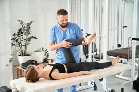


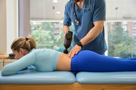


Explore the causes, treatment, and recovery timeline for a dislocated hip. Discover how physical therapy can facilitate your healing journey. For answers to any questions you may have, call Dr. Alexander Jimenez at 915-850-0900 or 915-412-6677